The role of inflammation in steady state and stress hematopoiesis


The response to systemic infection and tissue injury requires the rapid adaptation of hematopoietic stem cells (HSCs) in the bone marrow, which proliferate and divert their differentiation towards the myeloid lineage. Significant interest has emerged in understanding the signals that trigger this emergency hematopoietic program. However, the mechanisms that terminate this response of the HSCs and restore tissue homeostasis remain unknown. The clinical success of proteasome inhibitors, bortezomib, and E3 ubiquitin ligase glues for the treatment of hematologic diseases has made the Ubiquitin pathway a bona fide target for cancer therapeutics. Thus, defining how novel E3 ligases function in the bone marrow and investigating their specific roles in normal and emergency hematopoiesis can lead to novel therapeutic interventions. We have demonstrated (Guillamot et al. Nature Immunology, 2019) that the E3 ubiquitin ligase Spop restrains the inflammatory activation of HSCs. In the absence of Spop, systemic inflammation proceeds in an unresolved manner and the sustained response in the HSCs results in a lethal phenotype reminiscent of hyper-inflammatory syndrome. Our proteomic/biochemical studies demonstrated that Spop restricts inflammation by targeting the signal transducer Myd88 for proteasome-dependent degradation. Myd88 accumulation in conjunction with an inflammatory stimulus leads to Myddosome formation, the hyper-phosphorylation of the Irak4 kinase and activation of a number of transcription factor pathways (NF-kB, Jun, Pu.1, Cebpb). Our current studies try to: a) define the transcriptional and chromatin landscape changes imposed during initiation and Spop-regulated termination of emergency hematopoiesis, and b) the role of the Myddosome assembly, signaling and termination in the sensing of inflammation signals by the HSC. We are also focusing on the role of novel regulators of stress hematopoiesis in the aging of blood stem and progenitor cells.

Applying single-cell sequencing approaches to characterize the healthy and malignant bone marrow microenvironment


Leukemia is a clonal hematopoietic neoplasm characterized by the proliferation and accumulation of lymphoid or myeloid progenitor cells throughout the bone marrow. Extensive genomic characterization uncovered multiple candidates for targeted therapy. Unfortunately, most targeted therapies fail to elicit prolonged disease remission due to the emergence of pre-existent or de novo therapy-resistant leukemic clones. There is compelling emerging evidence that cell non-autonomous contributions to leukemia play a pivotal role in disease development, propagation, and maintenance. One has to consider the various cellular components of the bone marrow microenvironment that form among them a network of molecular interactions. This array of cell types includes immune cells, adipocytes, bone-forming osteoblasts, mesenchymal stromal cells, and vascular endothelial cells. Numerous studies support a role of the microenvironment in maintenance of the leukemic cells as well as treatment resistance.

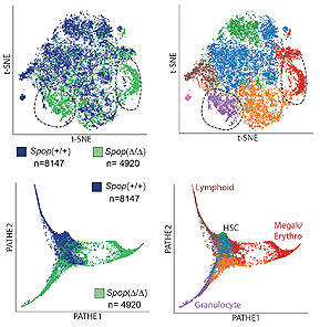
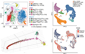
To better understand the function, heterogeneity, and the complexity of the bone marrow microenvironment in normal hematopoiesis and leukemia, we are using a combination of single-cell assays, including scRNA-Seq, CITE-Seq, and scATAC-Seq, in combination with computational methods to generate insights into this high-resolution biological data. Our work (Tikhonova, Dolgalev et al. Nature 2019) has characterized a previously unappreciated level of cellular heterogeneity within the bone marrow stromal niche, identified novel cellular subsets, reconstructed developmental trajectories, and resolved cellular sources of pro-hematopoietic growth factors, chemokines, and membrane-bound ligands. Under conditions of stress, our studies revealed a transcriptional remodeling of these niche elements accompanying the myeloid skewing that characterizes emergency hematopoiesis. To investigate the role of the microenvironment in B cell acute lymphoblastic leukemia (B-ALL) progression and treatment evasion, we utilized single-cell approaches to generate a comprehensive map of the primary human bone marrow immune microenvironment throughout three distinct stages of the disease: diagnosis, remission and relapse (Witkowski, Dolgalev et al. Cancer Cell 2020). We showed extensive re-modeling of the immune microenvironment composition throughout the course of conventional chemotherapy and uncover a role for leukemia-associated non-classical monocytes in promoting B-ALL pathogenesis in vivo. We took advantage of CITE-seq to expand the single-cell measurements beyond the transcriptome and simultaneously profile surface proteins, further validating our findings. Out studies provide a greater understanding of the potential extrinsic regulators of B-ALL survival and may highlight previously unknown environmental factors influencing immune-based treatment approaches to high-risk B-ALL.

Molecular Mechanisms of Drug Resistance in Acute Leukemia


Acute Myeloid Leukemia (AML) is a hematopoietic neoplasm associated with poor prognosis and high mortality, with an overall five-year survival rate of less than 30%. For patients with AML, standard induction regiments often fail to achieve the complete remission and disease relapse is often fatal, highlighting the need for novel efficient treatments. A greater understanding of the disease pathophysiology has led to the U.S. Food and Drug Administration (FDA) approval of eight new targeted therapies for the treatment of AML, since 2017, including the BCL-2 inhibitor venetoclax. Despite the favorable outcomes of AML patients treated with venetoclax in combination with hypomethylating agents or low-dose cytarabine, resistance to venetoclax ensues rapidly. Taking advantage of the CRISPR technology, our lab performed unbiased genome-wide screens to uncover the molecular mechanisms of resistance acquisition, as well as synergies in venetoclax treatment of human AML. Using this strategy, we identified loss of the tumor suppressor TP53 and depletion of the proapoptotic genes BAX and PMAIP (gene encoding for NOXA) as major modes conferring venetoclax resistance. In addition, we demonstrated that ablation of genes involved in mitochondrial organization and structure acts synergistically with BCL-2 inhibition. Transcriptomics, electron-microscopy and biochemical experiments revealed that mitochondrial shape adaptations contribute to the gain of venetoclax resistance. Our studies identified CLPB, a mitochondrial chaperonin, to be upregulated in human AML and further induced upon acquisition of venetoclax resistance. Genetic targeting of CLPB promotes apoptosis by inducing cristae remodeling and mitochondrial stress responses, thus sensitizing AML cells to venetoclax. Our work, published in 2019 at Cancer Discovery (Chen*, Glytsou* et al) suggests that targeting mitochondrial structure could be a promising strategy to overcome Venetoclax resistance in patients with AML. Further studies in the laboratory will focus on investigating the precise function of CLPB in normal hematopoiesis and leukemogenesis and its use as a potential therapeutic target. Moreover, additional CRISPR-based screens will be performed to shed light on liabilities and synthetic lethality of newly-developed targeted drugs for the treatment of distinct types of acute leukemia.

RNA-binding proteins (RBPs) in human cancer


RNA-binding proteins (RBPs) regulate many aspects of transcription and translation and are frequently dysregulated in cancers. Despite recent progress in uncovering essential RBPs in human leukemia maintenance, the promise of targeting RBPs therapeutically is still limited by a lack of systematic evaluation for required RBPs in cancer. Here, we have functionally dissected 490 classical RBPs in AML (and also T-ALL, melanoma and lung cancer) using a CRISPR (clustered regularly interspaced short palindromic repeat)-based domain targeting system. This uncovered a network of upregulated RBPs that are preferentially required for leukemia growth, including the novel splicing factor, RBM39. We have demonstrated that genetic or pharmacologic targeting of RBM39 consequently resulted in repressed cassette exon inclusion and promoted intron retention within mRNAs encoding HOXA9 targets as well as in other RBPs preferentially required for AML survival. Given the mechanistic role of RBM39 in splicing and the requirement of the DCAF15 adapter protein for anti-cancer sulfonamide activity, we identified that both the presence of spliceosomal gene mutations and levels of DCAF15 expression are important predictors of response to sulfonamides. We have also provided mechanistic support for expanded use of sulfonamides in clinical trials, as it identifies RBM39 as a key non-oncogenic addiction in AML, describes its mechanism of action, and offers valuable potential biomarkers and genetic predictors of response thereby presenting a novel strategy for treatment of AML patients. Future studies in the laboratory will focus on understanding the biological function of RBM39 in normal hematopoiesis and its transition into leukemia, as well understanding the role of other RBPs in cancers. Also, our CRISPR screens have identified a number of additional cancer type specific RBPs with key roles in a number of human cancers. We will focus on selected RBPs and aim to both understand their function and therapeutically target them using small molecules.

The role of TET proteins in hematopoietic malignancy


Recent studies have revealed a new layer of epigenetic regulation in stem cells and differentiated cell types whereby methyl groups at the 5-position of cytosine bases (5mC) can be modified by a novel family of α-keto-glutarate (α-KG)-dependent enzymes: Ten-Eleven-Translocation (TET) proteins, TET1, 2 and 3. All three TET proteins catalyze the conversion of 5mC to 5-hydroxymethylcytosine (5hmC), a DNA modification that is shown to be enriched at enhancers, promoters and gene bodies of actively expressed genes. The presence of 5hmC has been proposed by several studies to contribute to both passive and active DNA demethylation in the mammalian genome. Decreased expression of TET proteins and loss of 5hmC is a hallmark of many cancers, suggesting a critical role for the maintenance of this epigenetic modification in normal cellular function. The TET gene family was first identified because of the involvement of TET1 as a fusion partner of MLL in acute myeloid leukemia (AML). Studies in large cohorts of myeloid dysplastic syndrome (MDS) and AML patients revealed that somatic mutations in TET2 are found in 10 – 50% of cases. Our lab and other groups have generated knockout mouse models of Tet2 to confirm that loss of function promotes aberrant stem cell self-renewal and hematopoietic malignancy. In addition, we have found that Tet1 deletion in mice promotes lymphomagenesis. These studies highlight the non-redundant role of TET proteins to act as tumor suppressors of hematopoietic malignancy. Our current goal is to determine how modulation of the levels of 5mC and 5hmC predispose hematopoietic stem and progenitor cell to transformation. We are currently modeling the role of TET proteins in B, T and myeloid malignancy using conditional deletion and transgenic RNAi mouse models for inducible and reversible loss-of-function studies. Our goal is to identify novel and potential therapeutic targets in stem, progenitor and mature cells of TET proteins that regulate hematopoietic transformation and maintenance of aberrant DNA methylation.